
Fig. 5. Mode-of-action annotation of marine-derived natural products by FUSION mapping.
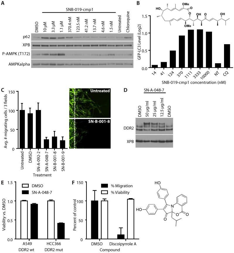
(A) Effect of SNB-019-cmp1 on autophagy and AMPK phosphorylation as measured by Western blot (A) or flow cytometry (B) of U2OS GFP-LC3 cells treated with the indicated concentrations of SNB-019-cmp1, chloroquine (50 μM), DMSO (0.1%), or untreated for two hours. Data are representative of five experiments. (B) Effect of SNB-019-cmp1 on autophagy as measured by the abundance of GFP-LC3 measured by flow cytometry in U2OS cells exposed to the indicated concentrations of SNB-019-cmp1, chloroquine (50 μM), or untreated (NT). Mean + SEM of 20,000 biological replicates. The structure of SNB-091-cmp1 is that of bafilomycin D (C) The effect of the indicated compounds (60 μg/ml) on human BR5 fibroblast migration out of a three-dimensional collagen plug induced by PDGF (50 ng/ml). Mean + SEM, n = 3 biological replicates. Right panels are representative images. Dotted lines indicate the boundary of the collagen plug. Cells were stained for filamentous actin. (D) Western blot for DDR2 in BR5 cells exposed to the indicated concentrations off SN-A-048-7. Data are representative of three experiments. (E) Normalized cell viability of A549 and DDR2-mutant HCC366 cells. Viability was measured using Cell TiterGlo. Data are mean + SEM of triplicate wells. These results are representative of five experiments. (F) The effect of discoipyrrole A (135 μM) or DMSO (0.1%) on the migration or viability of BR5 cells. Results were normalized to DMSO-treated cells. Mean + SEM. n = 3 experiments. The structure of SN-A-048-7 is that of discoipyrrole A.