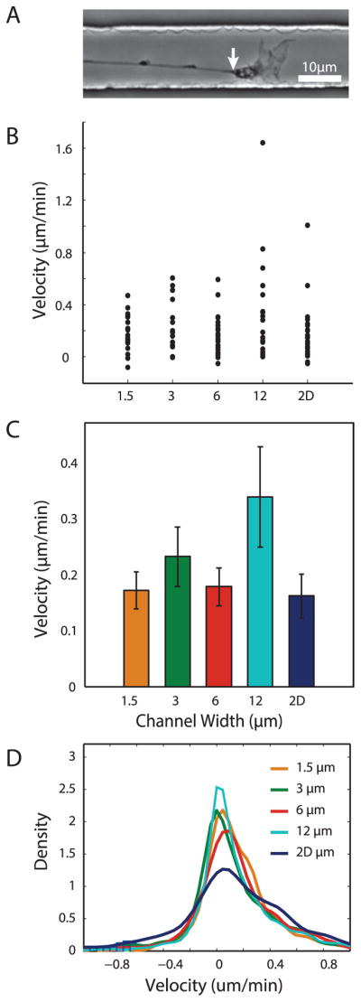
Figure 3.

Growth cone velocity is not affected global variations in channel width. (A) Representative brightfield image indicating wrist (white arrow) of growth cone used as marker for position. (B,C) Distribution and average of growth cone velocities based on total displacement in each group. N=19, 15, 22, 19, and 28 for 1.5μm, 3μm, 6μm, 12μm, and 2D, respectively. (D) Smoothed histogram of instantaneous velocities from all growth cone positions.
