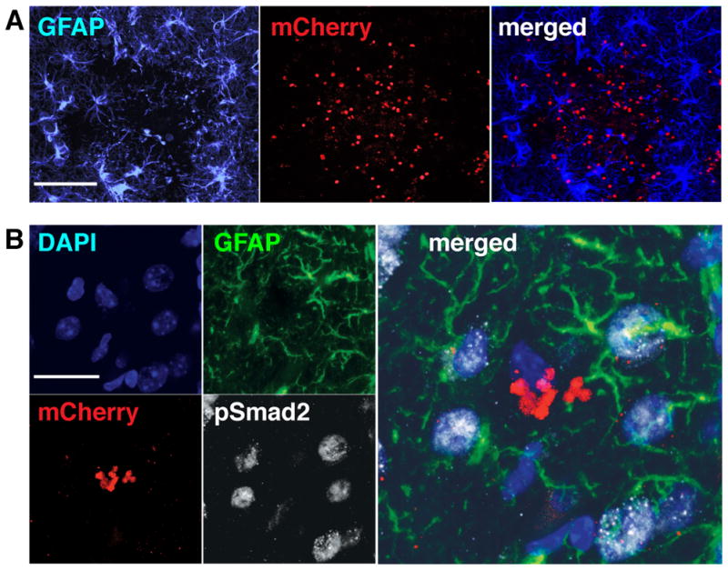
Figure 1. TGFβ signaling is activated in brain cells, including astrocytes during CNS toxoplasmosis.

Brain sections from wildtype mice infected with mCherry-expressing Toxoplasma were stained as indicated and then examined by confocal microscopy. A. Representative images showing astrocytes that immunostain for GFAP+ (left panel, blue), mCherry+ parasites (middle panel, red), and the merged image (right panel). Scale bar, 200 μm. B. Representative images of pSmad2 and GFAP co-immunostaining in areas surrounding mCherry+ Toxoplasma infiltrates. Panels: DAPI-stained nuclei (upper left panel, blue), GFAP+ astrocytes (upper right panel, green), mCherry+ parasites (lower left panel, red), pSmad2 staining (lower right panel, white), and merge of all 4 channels (right panel). Arrowheads highlight nuclei associated with the GFAP+ astrocytes seen in these images. Scale bar, 20 μm.
