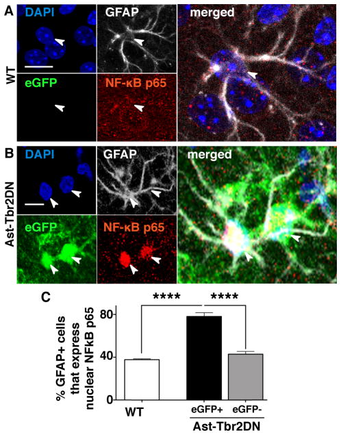
Figure 6. Lack of astrocytic TGFβ signaling increases NF-κB pathway activation in eGFP+ astrocytes.
A., B. Representative images of 2 wpi wildtype (WT) (A) and Ast-Tbr2DN (B) brain sections stained as indicated. Arrowheads denote the nuclei of the associated GFAP+ cells. C. Quantification of % of GFAP+ astrocytes that immunostain for nuclear NF-κB p65 in wildtype and Ast-Tbr2DN mice. Note that in Ast-Tbr2DN mice the GFAP+ cells are subdivided into GFAP+/eGFP+ and GFAP+/eGFP− cells. Scale bars, 20μm. N=6 mice per genotype. Bars, mean ± SEM. ****P<0.0001, 1-way ANOVA with Bonferroni correction for multiple comparisons.

