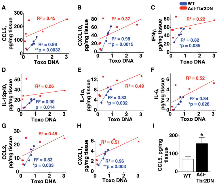
Figure 7. Inhibition of astrocytic TGFβ signaling leads to loss of correlation of Th1 cytokine and chemokine levels to acute CNS Toxoplasma burden.
At 2 wpi, brain homogenates from Ast-Tbr2DN and wildtype (WT) mice were used to quantify multiple cytokines and chemokines via multiplex cytokine assay and Toxoplasma burden (measured as B1 gene DNA, normalized to GADPH control gene DNA and expressed as fold over wildtype mean) via quantitative-PCR. A–H. Linear regressions of the Toxoplasma burden plotted against the levels of T cell chemokines CCL5 (A) and CXCL10 (B); Th1 cytokines IFNγ(C), IL-12p40 (D), IL-1a (E) and IL-6 (F); and myeloid cell chemoattractants CCL2 (G) and CXCL1 (H). Blue circles, wildtype mice. Red squares, Ast-Tbr2DN mice. There is a statistically significant linear correlation between Toxoplasma load and Th1 cytokines and chemokines in wildtype controls (blue line), but it is lost in Ast-Tbr2DN mice (red line). I, Mean CCL5 levels were approximately twice normal in Ast-Tbr2DN mouse brains. N=6 mice per genotype. Bars, mean ± SEM. *P<0.05, Student’s t test.

