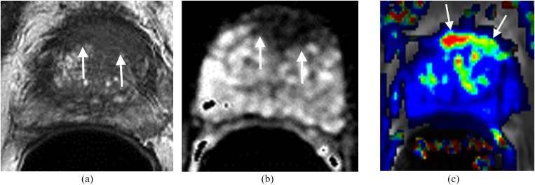
Figure 2.
Lesion in the anterior transitional zone (TZ). The patient had four prior negative transrectal ultrasound prostate biopsies and prostate-specific antigen of 17 ng ml−1. (a) Axial T2 weighted imaging shows a homogeneous low signal mass with indistinct margins (erased charcoal sign) (arrows) in the anterior aspect of the TZ. (b) Apparent diffusion coefficient shows the mass with low signal intensity (arrows). (c) Dynamic contrast-enhanced imaging demonstrates the lesion with rapid contrast wash-in and wash-out (arrows). MRI-guided biopsy confirmed prostate carcinoma Gleason score 9.

