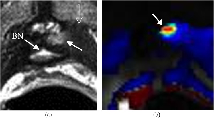
Figure 4.
Small recurrence in the left side of the prostate surgical bed. The patient's status is post radical prostatectomy with a recent elevation of prostate-specific antigen to 0.21 ng ml−1. (a) Axial T2 weighted imaging shows a small mass with slightly high T2 signal (solid arrow) relative to the adjacent muscle (open arrow) in the left side of the prostate surgical bed. Bladder neck (BN) is noted. (b) Dynamic contrast-enhanced imaging shows the lesion with rapid contrast wash-in and wash-out (arrow) consistent with focal recurrence.

