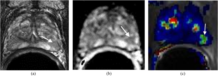
Figure 5.
Focal chronic prostatitis in the left side of the peripheral zone. The patient had negative transrectal ultrasound (TRUS) prostate biopsy with an elevation of prostate-specific antigen to 8.1 ng ml−1. (a) Axial T2 weighted imaging shows a mildly low T2 signal focus (arrow) in the left side of the peripheral zone. (b) Apparent diffusion coefficient (ADC) map shows the focus with mildly a decreased ADC value (arrow). (c) Dynamic contrast-enhanced imaging shows bilateral symmetric enhancement (arrow). MRI-guided prostate biopsy confirmed chronic prostatitis.

