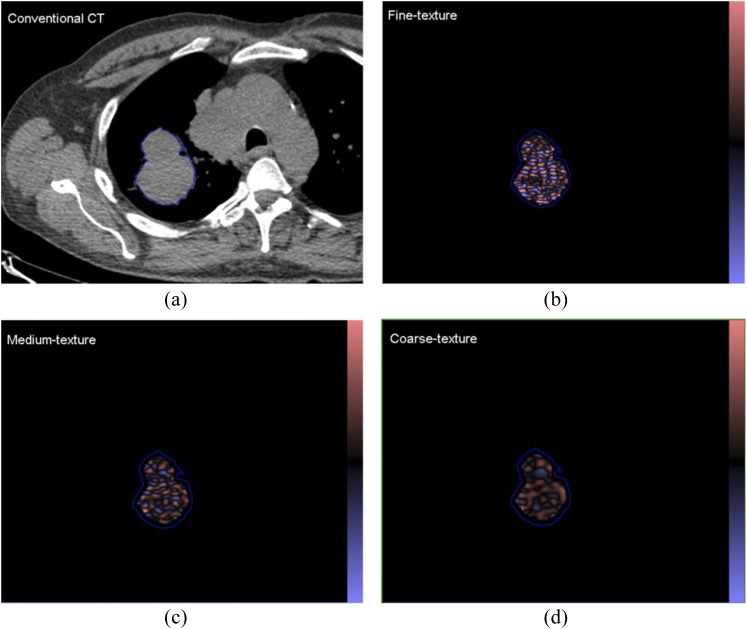
Figure 2.
(a) A conventional CT (from a positron emission tomography/CT) image of a patient with a lung lesion and (b–d) corresponding images selectively displaying fine, medium and coarse texture obtained from TexRAD CT texture analysis (image heterogeneity) commercial research software (www.texrad.com, Radstock, UK). Images should be viewed in the online format.

