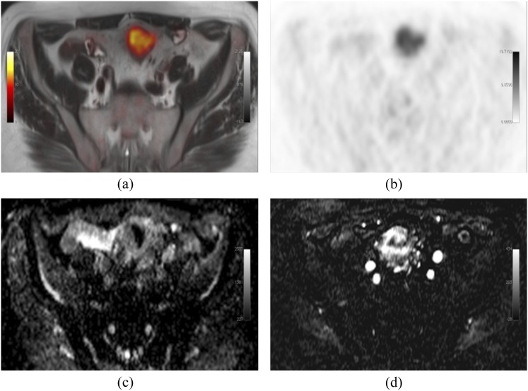
Figure 4.
Produced from an imaging unit at the Institute of Nuclear Medicine, University College London, UK. Simultaneous 18F-fludeoxyglucose–positron emission tomography (PET)/MRI-acquired image of a patient with a sigmoid tumour. Fused axial T2 and PET (a), PET alone (b), MRI apparent diffusion coefficient map (c) and representative subtracted image from a dynamic contrast-enhanced MRI series (d); showing increased metabolism, cellularity and vascularity. Images should be viewed in the online format.

