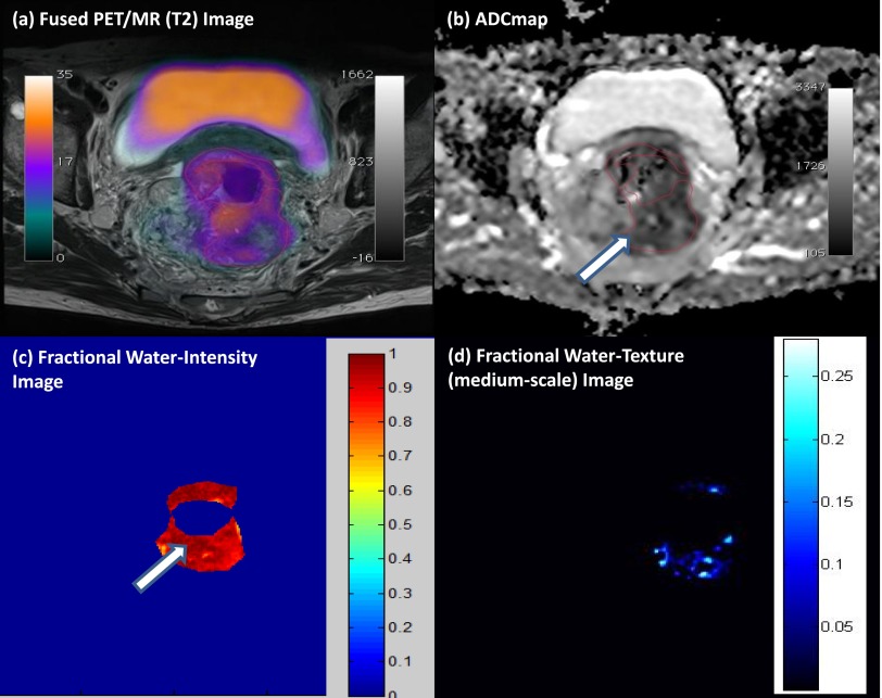
Figure 5.
Multiparametric positron emission tomography (PET)/MRI of a rectal cancer. (a) High 18F-fludeoxyglucose uptake on fused PET/T2 MRI, with (b) a correspondingly patchy reduced apparent diffusion coefficient (ADC) in keeping with pockets of high cellularity within the tumour and (c) a fractional water image derived from source fat and water Dixon images of the same tumour confirms that areas of increased cellularity correlate with relatively increased water content (white arrows). (d) Application of a medium coarse textural filter highlights 3- to 4-mm bright objects on the fractional water image (medium texture map). Images should be viewed in the online format. (www.texrad.com, Radstock, UK.)

