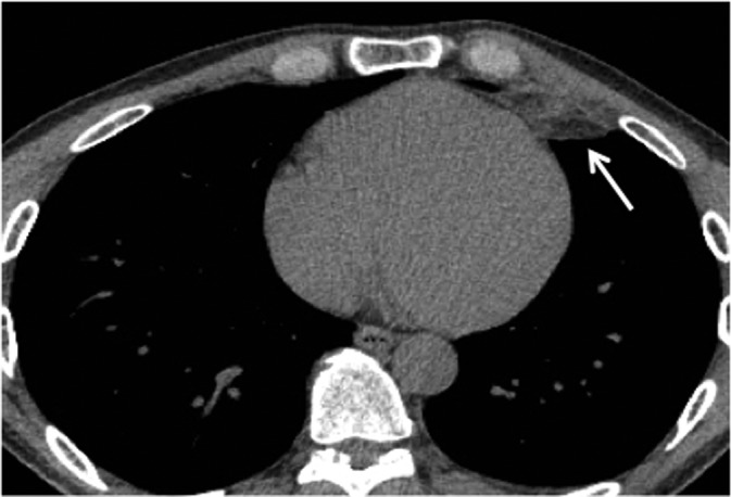
Figure 2.

A 24-year-old male with 3-day left pleuritic chest pain and epipericardial fat necrosis findings. The axial view of an enhanced chest CT scan displays a small round soft-tissue attenuation lesion with mild stranding (arrow) close to the diaphragmatic pleural surface in the epipericardial fat.
