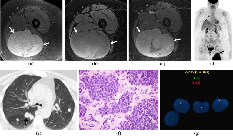
Figure 2.
A 63-year-old woman with extraskeletal Ewing sarcoma of the thigh. (a–c) Axial fat-suppressed T2 weighted, pre- and post-gadolinium fat-suppressed T1 weighted MRI images demonstrate a heterogeneously hyperintense mass on T2 weighted images (arrows), which is heterogeneously isointense on T1 weighted images with heterogeneous enhancement. (d) Coronal maximum intensity projection image of fluorine-18 fludeoxyglucose (18F-FDG) positron emission tomography/CT FDG uptake (maximum standardized uptake value of 12) in the right thigh mass (arrow) and multiple 18F-FDG-avid pulmonary metastases (arrowheads). (e) Axial CT image of the chest confirms the pulmonary metastases (arrow). (f). Haematoxylin and eosin staining (original magnification ×600) of the biopsy specimen of the tumour demonstrates sheets of uniform rounded cells with scant cytoplasm. (g). Fluorescence in situ hybridization analysis using break-apart probes directed against the 5′ and 3′ ends of the EWSR1 gene shows EWSR1 gene rearrangement.

