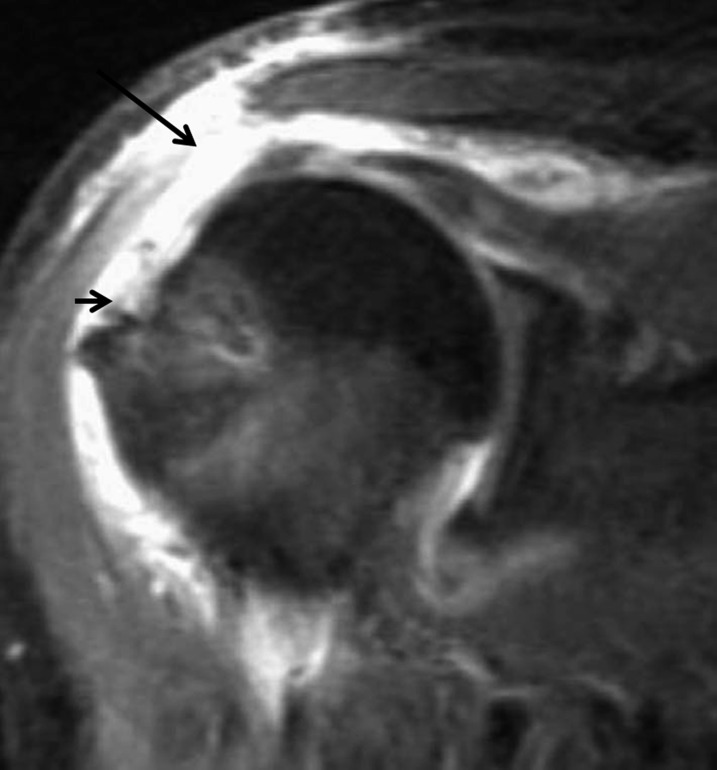
Figure 15.
Fat-suppressed intermediate-weighted (repetition time/echo time, 1883/41 ms) coronal view of an elderly female with continued weakness that shows complete detachment of the deltoid at the acromial attachment site with a fluid-filled defect (large arrow). There is also a recurrent full-thickness tear of the supraspinatus (small arrow).

