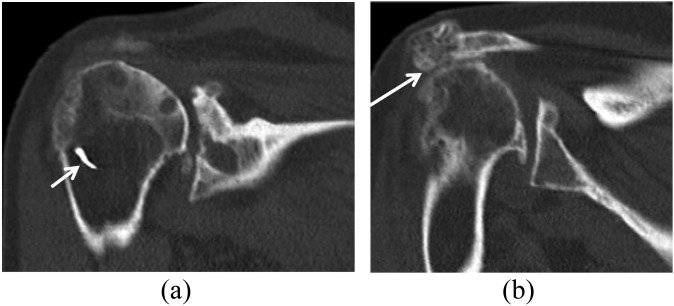
Figure 4.
Radiographic findings of a 75-year-old female with a history of subacromial decompression and a failed rotator cuff repair. (a) Coronal CT image showing the position of the metallic suture anchor within the bone distal to the greater tuberosity (arrow) and superior migration of the humerus and glenohumeral joint osteoarthrosis, with extensive cyst formation along the articular surface of the acromion and humeral head. (b) Coronal CT image shows reformation of the lateral acromial osteophyte (arrow).

