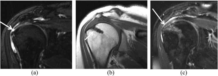
Figure 6.
Common soft-tissue findings on MRI of a middle-aged male before and after rotator cuff repair and subacromial decompression. (a) Pre-operative fat-suppressed T2 weighted (repetition time/echo time, 3380/90 ms) coronal image showing a small full-thickness tear of the supraspinatus tendon (arrow). (b) After rotator cuff repair with subacromial decompression, T1 weighted (repetition time/echo time, 530/10 ms) coronal view shows a suture anchor (arrow) as a susceptibility artefact related to the presence of a bioabsorbable anchor. (c) Fat-suppressed T2 weighted (repetition time/echo time, 3380/90 ms) coronal image shows intermediate signal in the tendon, a common and expected finding after surgery (arrow).

