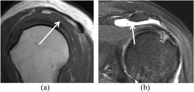
Figure 7.
A clinically asymptomatic 54-year-old female after acromioplasty and rotator cuff repair. (a) T1 weighted (repetition time/echo time, 530/10 ms) sagittal image shows a diminutive undersurface of the acromion related to acromioplasty (arrow). An excessively thinned acromion is susceptible to fracture and can be further evaluated if necessary with CT. (b) Fat-suppressed intermediate-weighted (repetition time/echo time, 3200/45 ms) coronal image obtained after surgery shows fluid in the subacromial–subdeltoid bursa (arrow).

