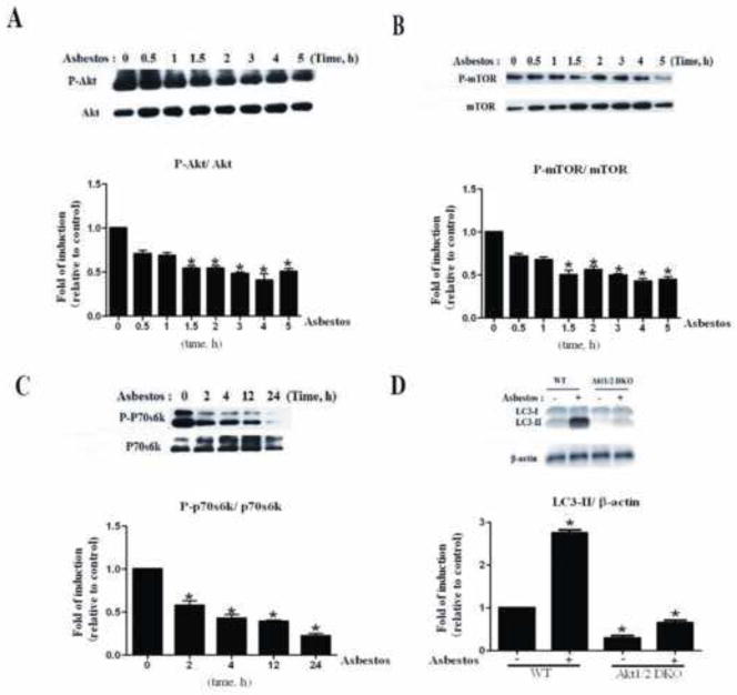
Fig. 3. Chrysotile asbestos treatment induced autophagy in A549 cells and MEFs via the PI3K/AKT/mTOR pathway.
A549 cells were treated with chrysotile asbestos for the indicated time (0–24 h). The expression levels of the indicated proteins were analyzed by immunoblotting. (A–C) Western blot assays were used to examine the total and phosphorylated protein levels of AKT, mTOR, and P70S6K. Densitometry signals were normalized to those of total protein, then normalized to those of untreated A549 cells (100%). (D) Western blot of LC3 in WT and AKT (DKO) MEF cells. The ratio of LC3II/β-actin was determined. The intensity of these protein signals obtained was quantified using Image J software from three replicate experiments.

