. 2013 Jun-Aug;39(4):513–517. doi: 10.1590/S1806-37132013000400016

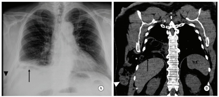
Figure 1. In A, posteroanterior chest radiograph. The arrow demonstrates opacification at the base of the right hemithorax, interspersed with some gas images. The arrowhead indicates soft tissue swelling in the periphery of the right lateral chest wall, with some gas images similar to those observed at the base of the right hemithorax. In B, coronal reconstruction of a noncontrast CT scan of the chest showing herniation of abdominal content into the base of the right hemithorax through the diaphragm (arrow), as well as through the eighth intercostal space (asterisk), headed toward the subcutaneous tissue of the chest wall. Note the presence of thin loops in the subcutaneous tissue of the chest wall (arrowhead).

Figure 1