. 2013 Jun-Aug;39(4):513–517. doi: 10.1590/S1806-37132013000400016

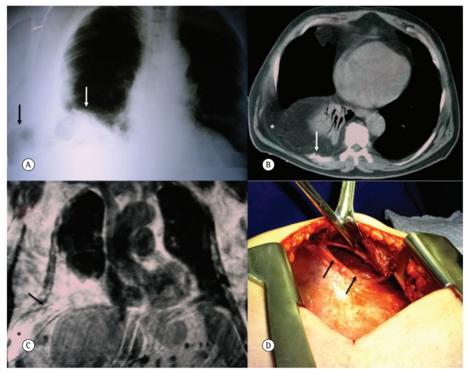
Figure 3. In A, posteroanterior chest radiograph demonstrating opacification at the base of the right hemithorax, superiorly displacing the lung parenchyma (white arrow). Note the widening of the eighth intercostal space and the presence of a structure with gas density, similar to an intestinal loop, occupying the soft tissues of the adjacent chest wall (black arrow). In B, axial CT image of the chest, where it is possible to observe the passage of epiploic fat into the base of the right hemithorax and subcutaneous tissue of the chest wall (asterisk). Note the presence of a fracture of the ninth rib (white arrow). In C, coronal T1-weighted non-fat-saturated magnetic resonance image demonstrating the presence of a large amount of abdominal fat herniated into the base of the right hemithorax. Note the discontinuity of the intercostal muscles (black arrow), through which the content herniates into the subcutaneous tissue of the chest wall. In D, photograph showing the main surgical finding: a rent in the anterolateral portion of the diaphragm.

Figure 3