
Figure 2.
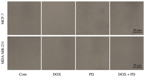
Morphological changes in breast cancer cells after DOX, PD, and DOX + PD treatments. MCF-7 and MDA-MB-231 cells were treated with 5 μM DOX, 10 μM PD, or 5 μM DOX + 10 μM PD. After 24 h of treatment, the cell morphological changes were observed with an inverted-phase contrast microscope.