
Figure 4.
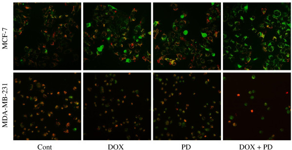
JC-1 staining of breast cancer cells after DOX, PD, and DOX + PD treatments. MCF-7 and MDA-MB-231 cells were treated with 5 μM DOX, 10 μM PD, or 5 μM DOX + 10 μM PD. After 4 h of treatment, JC-1 fluorescence was utilized to study the mitochondrial membrane potential change. The cells were observed with a fluorescent microscope, and images were obtained with an Axiovert 200 fluorescent inverted microscope and an AxioCam HRc CCD camera.