Figure 5.

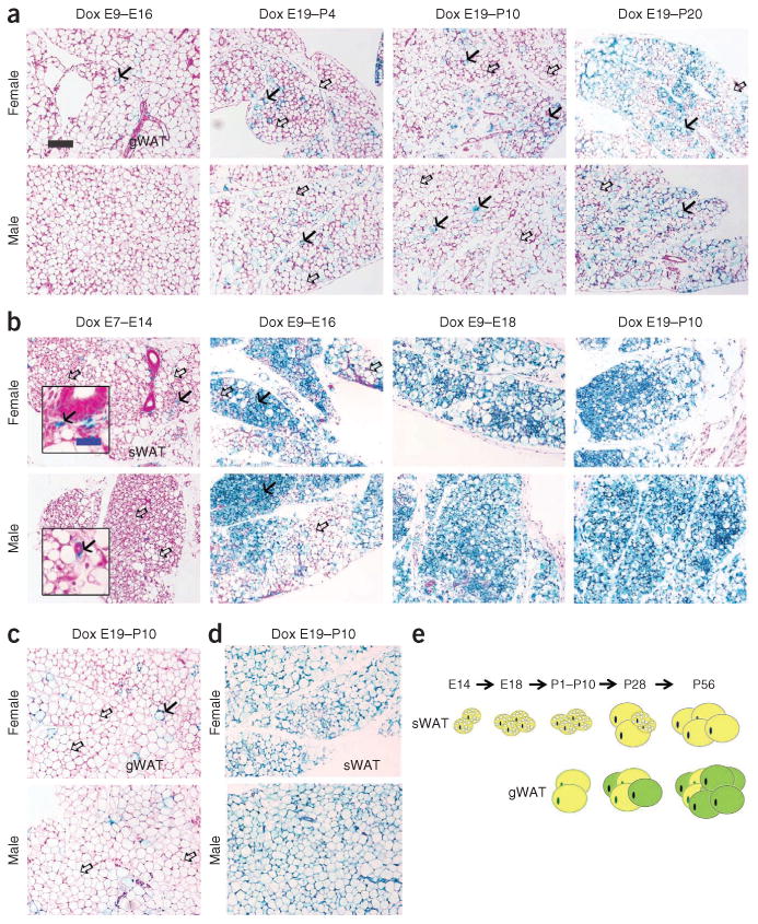
Development of epididymal and subcutaneous adipocytes during embryonic and postnatal development. (a-d) Results from AdipoChaser mice that were on doxycycline diet for the indicated number of days during embryonic and postnatal development that were thereafter kept on chow diet. (a) Representative β-gal staining of gonadal WAT (gWAT) from 28-day-old female mice (top) and male littermates (bottom). The mothers of these mice were on doxycycline diet during E9-E16, E19-P4, E19-P10 or E19-P20, as indicated. (b) Representative β-gal staining of sWAT from 28 day-old-female (top) and male (bottom) mice. The mothers of these mice were on doxycycline diet during E7-E14, E9-E16, E9-E18 or E19-P10, as indicated. (c,d) Representative (3-gal staining of gWAT of 56-day-old female (c, top) and male (c, bottom) mice and of sWAT from 56-day-old female (d, top) and male (d, bottom) mice. The mothers of these mice were on doxycycline diet during E19-P10. Solid arrows (a–c), LacZ-positive cells; open arrows (a–c), LacZ-negative cells. Scale bar (black, shown in a, applies to a–d). 200 μm; (blue, shown in b, applies to the insets in b), 50 μm. For a–d, n = 2 mice per group. (e) Schematic model of the development of gWAT and sWAT. Adipocytes in the gWAT are differentiated postnatally between birth and sexual maturation, whereas all the adipocytes in the subcutaneous adipose tissue start to differentiate between E14 and E18, but the differentiation takes much longer and finishes postnatally. Yellow adipocytes represent adipocytes differentiated before birth, and green adipocytes represent adipocytes differentiated postnatally.
