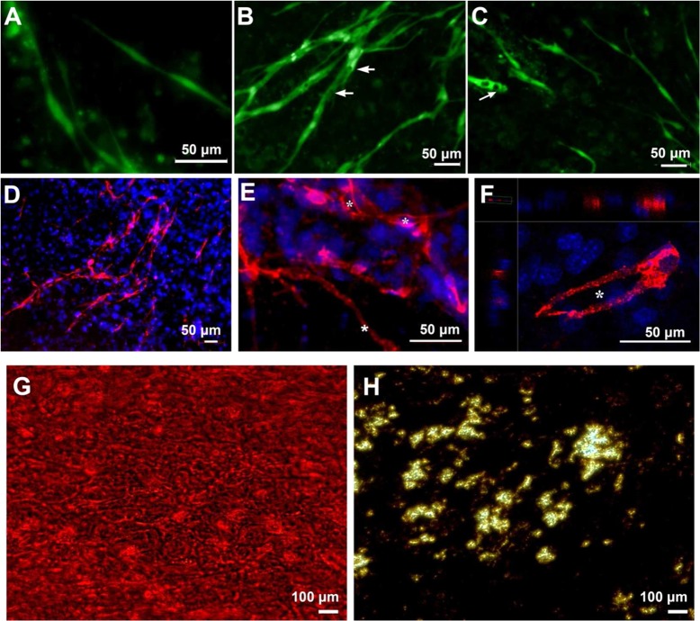
Figure 2.
HUVECs on an undifferentiated hMSCs sheet formed numerous networks. Networks started at day 3 (A), and elongated to form many lumens at day 5 (B) and day 7 (C). Arrows indicate lumens. Immunofluorescent staining images of CD31 show several networks on the hMSCs sheet at day 7 (D, 10× magnification; E, 20× magnification); 3D-reconstructed confocal images display lumen formation (F). Asterisks show the lumens. Alizarin red staining (G) and vov Kossa staining (H) show the mineralized matrix of osteogenic hMSC cell sheet.

