Figure 3.
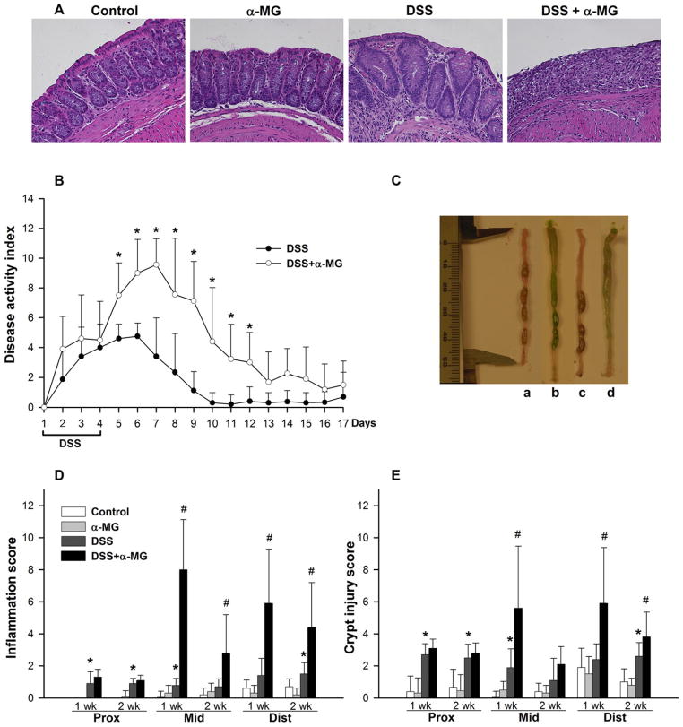
(A) Hematoxylin and eosin staining in distal colon from mice following 1-week recovery after administering dextran sulfate sodium (DSS). Magnification: 20×. (B) Dietary α-mangostin (α-MG) exacerbates disease activity index (*p < 0.05). (C) Increased fluid content in colonic lumen of mice fed AIN93G diet with α-MG; experimental groups: a, control; b, α-MG; c, DSS; d, DSS + α-MG. The green pigmentation in the colonic lumen of mice fed diet with α-MG is the dye added to the diet (see Methods). (D) Inflammation and (E) crypt injury scores in the mid and distal colon are greater in the DSS + α-MG group compared to DSS group after 1 and 2 weeks of recovery (*p < 0.05 against control; #p < 0.05 against DSS group). The data points represent the mean (±SD) of values from nine to ten mice per group.

