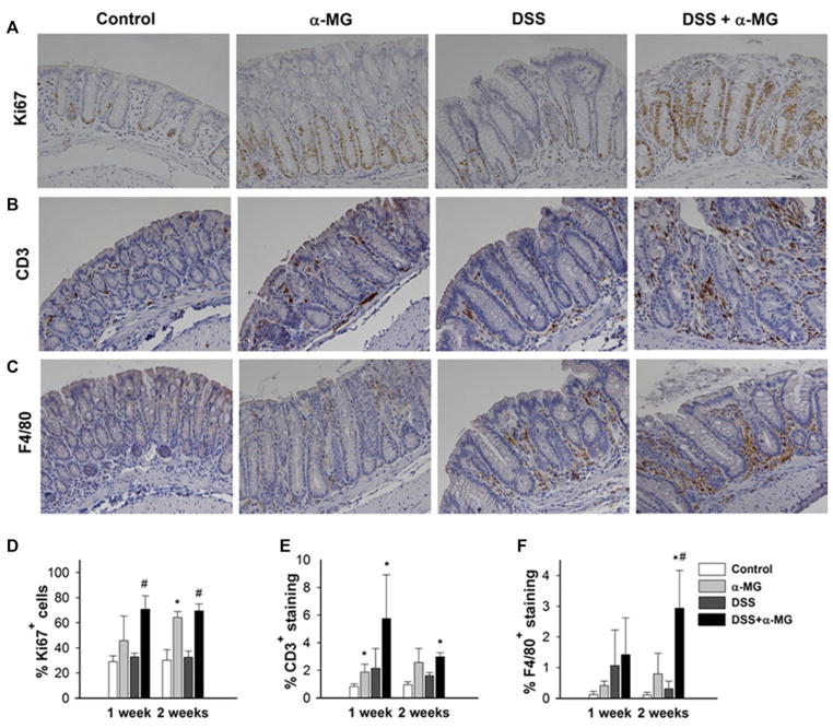
Figure 4.
Representative images of (A) Ki67, (B) CD3, and (C) F4/80 immunostaining in distal colon of mice 1 week after cessation of dextran sulfate sodium (DSS) treatment. Magnification: 20×. (D) Ki67+ immunostaining of colonic epithelial cells was increased in animals fed diet with α-mangostin (α-MG). (E) CD3+-stained tissue for T cells in the colonic lamina propria of mice fed diet with α-MG was greater than in the control group. (F) Significant macrophage infiltration, as determined by immunostaining for F4/80, in the DSS + α-MG 2 weeks after recovery. *p < 0.05 against control group; #p < 0.05 against DSS group). The data points represent the mean (±SD) of values from three mice per group.

