
Figure 4.
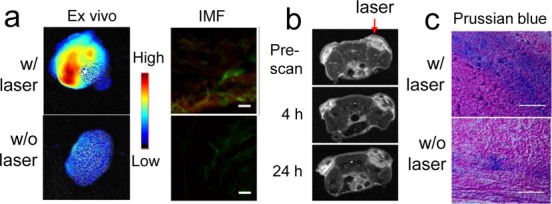
EPR enhancement with nanoparticles. (a) EPR enhancement effect with QDs. The study was performed in bilateral 4T1 tumor models. QDs were injected 5 min after the end of P-RFRT-mediated PDT, which was applied only to the left-side tumors. Ex vivo imaging was performed on dissected tumors 24 h after the QD injection. Compared to unirradiated tumors, irradiated tumors showed a 20.8-fold increase in tumor uptake (by ROI analysis). This was further confirmed by immunofluorescence (IMF) staining. Green, CD31, marks blood vessels; red, QDs. Scale bars, 100 μm. (b) EPR enhancement effect with IONPs (n = 3). The study was performed in bilateral 4T1 tumor models. IONPs were injected 5 min after the end of P-RFRT-mediated PDT, which was applied only to the right-side tumors. MR images were taken before and 4 and 24 h after the injection of IONPs. More significant signal drop was observed in the right-side, irradiated tumors. (c) Prussian blue staining on tumor samples from b. Correlated with the in vivo observations, more iron deposits were found in irradiated tumors. Scale bars, 100 μm.