
Figure 5.
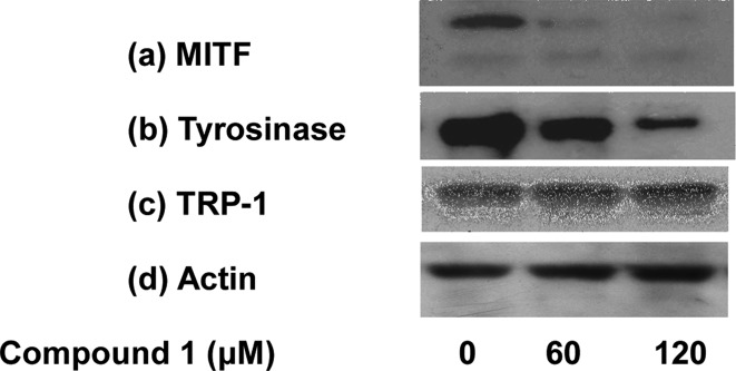
Expression of melanogenesis-related proteins in melan-a melanocytes after 1 treatment for 1 day in FSK-supplemented medium. Cell lysates were prepared from treated melanocytes following 1 treatment. The expressions of MITF (a), tyrosinase (b), and TRP-1 (c) were analyzed by Western blotting, and the expression of melanogenic proteins relative to actin is shown in panel (d).