Fig. 3.
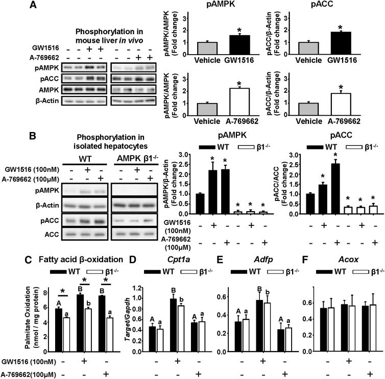
GW1516 increases AMPK and ACC phosphorylation, which is not required for FA oxidation. A: Eight- to 10-week-old Ldlr−/− mice fed a standard laboratory chow were fasted overnight, fed at 07:00 h for 2 h, and refasted at 09:00 h. Intraperitoneal injection of vehicle, GW1516 (3 mg/kg), or A-769662 (30 mg/kg) (n = 6/group) occurred at the beginning of the refasting period at 09:00 h. Immunoblots of AMPK and ACC in freeze-clamped liver lysates 90 min postinjection. Representative immunoblots with quantitations are shown. Asterisk (*) indicates significant difference between vehicle and treatment; Student’s paired t-test (P < 0.05). B–F: Primary hepatocytes isolated from WT and AMPKβ1−/− mice. Cells were incubated for 1 h with or without GW1516 or A-769662, and lysates were immunoblotted for phosphorylated AMPK and ACC (pAMPK, pACC) (B). Representative immunoblots with quantitations are shown. Asterisk (*) indicates significant difference versus WT control; Student’s paired t-test (P < 0.05). C: Isolated hepatocytes were treated with 0.5 mM palmitate (0.5 μCi/ml [14C]palmitate) for 4 h with or without GW1516 or A-769662 prior to determination of FA oxidation. In isolated hepatocytes treated with or without GW1516 or A-769662 for 6 h, mRNA abundances of Cpt1a (D), Adfp (E), and Acox (F) were measured by quantitative real-time PCR and normalized to Gapdh. Different uppercase letters indicate statistical significance among treatments in WT hepatocytes, different lowercase letters indicate statistical significance among treatments in β1−/− hepatocytes, and an asterisk (*) indicates statistical significance between WT and β1−/− within the same treatment group (P < 0.05); two-way ANOVA with post hoc Tukey’s test (P < 0.05). All data are presented as mean ± SEM (n = 3–4 from at least three independent experiments).

