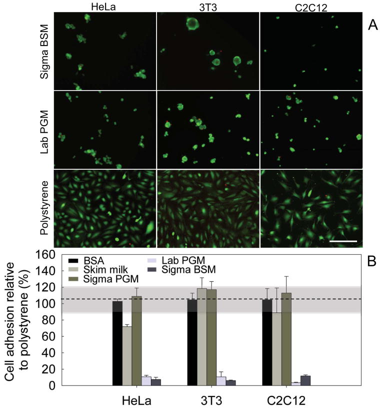
Figure 1. Mucin coatings are cytophobic.
(A) HeLa epithelial cells, 3T3 fibroblast cells and C2C12 myoblasts were seeded on a polystyrene surface, or on coatings generated from BSM (Sigma) or PGM (in-lab purified) and labeled with a live (green)/dead (red) stain. Scale bars: 200μm.” (B) Quantification of cell adhesion on mucin coatings generated with commercial BSM and PGM (Sigma BSM/PGM), in-lab purified PGM (Lab PGM), Skim milk, and BSA. The highlighted region between 90–120% is the standard deviation of the reference adhesion to polystyrene.

