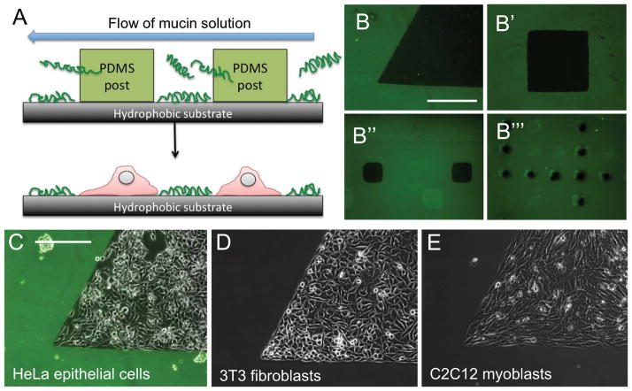
Figure 2. Mucin coatings for cell patterning.
(A) Mucin coatings were patterned using a microfluidic device with posts masking defined areas of the surface. (B) Patterns of mucin-free areas (in black) can be generated in different sizes and shapes. When seeded on the patterned surfaces, the epithelial cells (C), fibroblasts (D) and myoblasts (E) accumulated in the uncoated regions, avoiding the mucin coatings. Scale bars: 250μm.”

