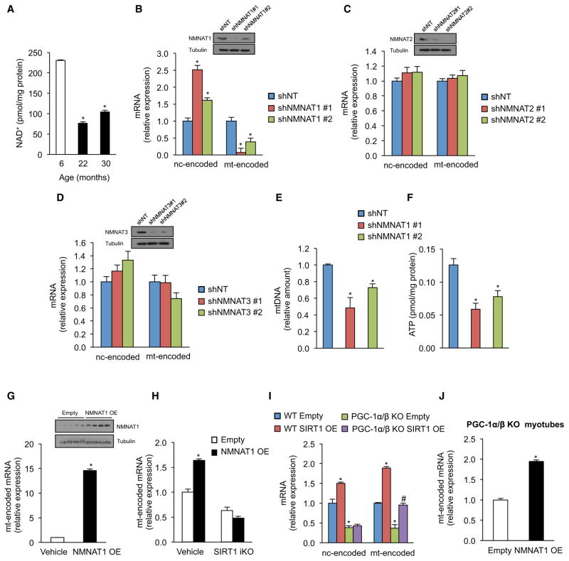
Figure 2. Nuclear NAD+ Levels Regulate Mitochondrial-Encoded Genes and Mitochondrial Homeostasis through SIRT1, Independently of PGC-1α/β.
(A) NAD+ levels in gastrocnemius of 6-, 22-, and 30-month-old mice (n = 5, *p < 0.05 versus 6-month-old mice).
(B–D) Expression of nuclear- and mitochondrially encoded genes in primary myoblasts transduced with NMNAT1 (B), NMNAT2 (C), NMNAT3 (D), or nontargeting shRNA (n = 4, *p < 0.05 versus shNT).
(E and F) Mitochondrial DNA content and (H) ATP content (I) in primary myoblasts transduced with NMNAT1 or nontargeting shRNA (n = 4, *p < 0.05 versus shNT).
(G) Expression of mitochondrially encoded genes in tibialis of 10- to 12-month-old mice overexpressing NMNAT1 compared to the contralateral tibialis muscle treated with vehicle (n = 4, *p < 0.05 versus vehicle).
(H) Expression of mitochondrially encoded genes in SIRT1 flox/flox Cre-ERT2 primary myoblasts treated with vehicle or OHT to induce SIRT1 excision infected with adenovirus overexpressing NMNAT1 or empty vector (n = 4, *p < 0.05 versus vehicle empty vector).
(I and J) Expression of nuclear- and mitochondrially encoded genes in WT and PGC-1α/β knockout myotubes treated with adenovirus overexpressing SIRT1 (I) or NMNAT1 (J) (n = 4, *p < 0.05 versus WT empty; #p < 0.05 versus PGC-1α/β KO empty).
Nuclear- and mitochondrially encoded genes were ND1, Cytb, COX1, ATP6 and NDUFS8, SDHb, Uqcrc1, COX5b, ATP5a1, respectively. Tissue samples are gastrocnemius muscle unless otherwise stated. Values are expressed as mean ± SEM. See also Figure S2.

