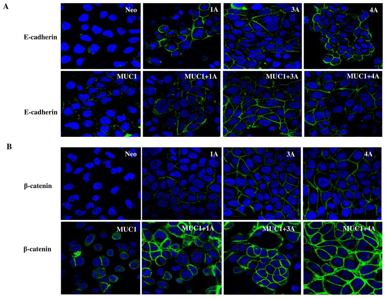
Fig. 5. Immunofluorescence of E-cadherin and β-catenin expression in control (neo) and MUC1 expressing (MUC1) S2-013 cells with and without re-expression of the indicated isoforms of p120 catenin.
(A) E-cadherin is not stabilized in the absence of p120 catenin in S2-013.Neo cells. Re-expression of p120 isoforms 1A, 3A, 4A in S2-013 cells restored E-cadherin expression. Green indicates E-cadherin staining, Blue are nuclei. (B) β-catenin expression was extremely low in control S2-013 cells that lacked p120 catenin. Expression of MUC1 alone or re-expression of p120 catenin isoforms enhanced slightly the levels of β-catenin at cell junctions. There was a dramatic increase in levels of junctional β-catenin in cells expressing MUC1 and any of the p120 catenin isoforms. β-catenin staining is shown as green and nuclei are blue.

