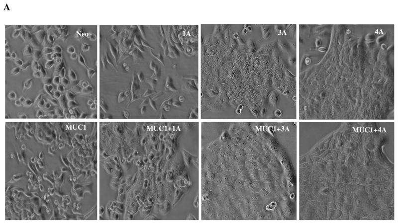
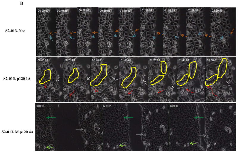
Fig. 6. Expression of different p120 catenin isoforms affects epithelial morphology and cell motility of S2-013 cells.
(A) Phase contrast photographs of control (neo) S2-013 cells and MUC1-expressing S2-013 cells with re-exepression of different p120 catenin isoforms. (B) Time lapse microscopy images showing three different types of cell motility that were produced upon expression of different p120 catenin isoforms (elapsed time indicated in each panel and arrows indicate position of individual cells in the monolayer at the different time points): control cells S2-013 neo showed weak and pliable cell-cell contacts that are maintained as a monolayer; dynamic changes of cell motility were seen in the S2-013 cells with p120 catenin 1A expression, which showed a high degree of motility in limited space but generally remained associated with other cells in the monolayer by highly pliable and exchangeable contacts; there was a high rate of organized and unified growth and motion in the direction of wound closure by S2-013 cells expressing MUC1 and p120 catenin 4A. Yellow circle shows dynamic features of movement of a group of cells. Red arrow indicates a cell migrating underneath the neighboring cells with greatly enhanced motility. Scale, 10 μm.


