
Figure 1.
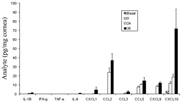
Expression of inflammatory cytokines and chemokines in the cornea of HSV-1 infected mice. C57BL/6 female mice (n=6/timepoint) were left alone (basal) or scarified (0) and infected with HSV-1 (McKrae strain, 1000 plaque forming units (PFU)/eye. Twenty-four to thirty-six hours postinfection, the mice were euthanized, perfused, and the cornea was removed and homogenized in a buffer containing a cocktail of protease inhibitors. The supernatant was clarified (10,000 × g, 5 min) and assayed for cytokine/chemokine content by ELISA. Bars represents mean ± SEM for each analyte under measure.