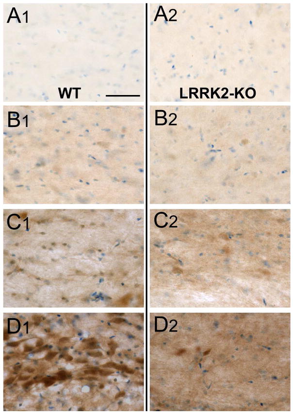
Figure 5. Titration of anti-LRRK2 antibodies in the rat SNpc.
SNpc from adult WT (left panels, labeled subpanels 1) and LRRK2 KO rats (right panels, labeled subpanels 2), with increasing concentrations of primary antibody (N241A/34) at A) 1 μg/ml (standard concentration used in all other experiments), B) 2.5 μg/ml C) 13 μg/ml and D) 40 μg/ml, together with secondary antibody held at 1 μg/ml for all panels. Nissl stain was provided to aid in the identification of the SNpc. The lateral SNpc is shown for all panels, with both LRRK2 WT and LRRK2 KO sections processed in parallel. Scale bar is 40 μm for all panels.

