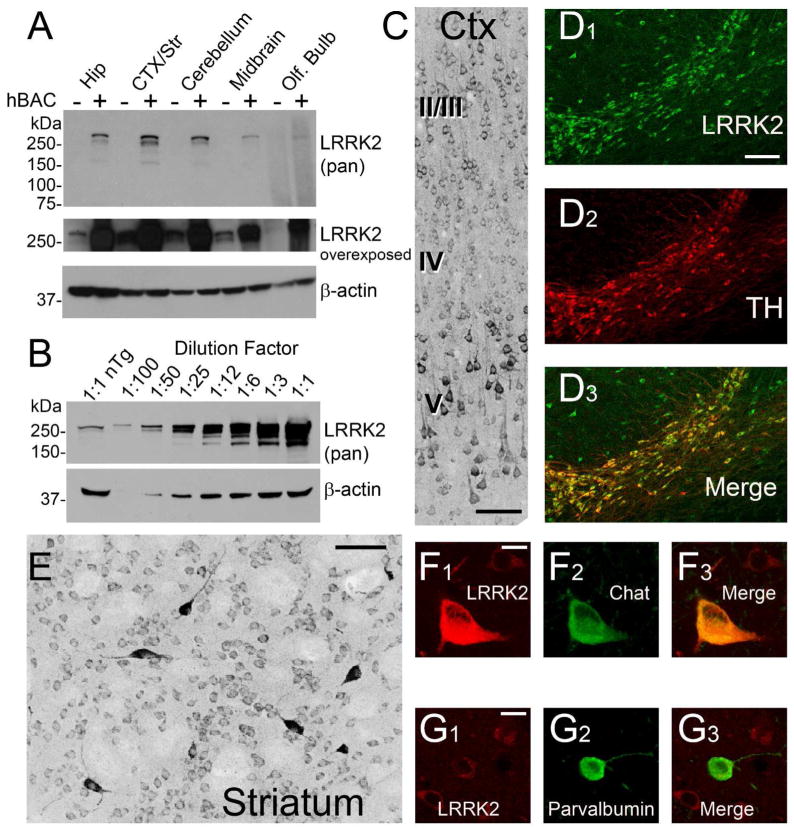
Figure 7. Aberrant LRRK2 Distribution Caused by Expression of Human LRRK2 BAC Constructs in Rats.
A) Western blot analysis with antibody N241 demonstrating the level of LRRK2 overexpression in the indicated brain region, as dissected from fresh rat brain tissue from either nontransgenic or LRRK2-G2019S human-BAC transgenic rats. Equivalent protein concentrations determined by BCA assay were loaded between lanes (and verified by actin signal), matched by region, between wild-type and transgenic rats. B) Forebrain lysate from nontransgenic rats was loaded in lane 1 and increasing concentrations of LRRK2-G2019S transgenic rat forebrain lysates were loaded in subsequent lanes. LRRK2 is overexpressed between 25 and 50 fold in the transgenic rats. C)The pan-reactive LRRK2 antibody N241A/34 was used to detect LRRK2 in LRRK2-G2019S hBAC positive rats in the motor cortex (scale bar is 50 μm), and D) SNpc, by fluorescent co-label with TH (scale bar is 0.1 mm). E) A striosome in the anterior striatum of transgenic rats identified by LRRK2, with intensely positive LRRK2 interneurons inside the striosome (scale bar is 50 μm. F,G) Co-localization of LRRK2 with cholinergic interneurons, but weak expression in parvalbumin-positive interneurons (scale bars are 10 μm). A magenta-green version of the images is available as Supplemental Figure 5.

