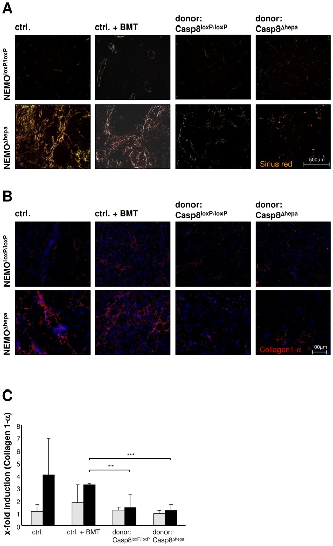
Figure 3. Reduced fibrogenesis in hepatocyte transplanted NEMOΔhepa mice.
(A) Analysis of Collagen accumulation in transplanted mice via Sirius red staining under polarized light. NEMOΔhepa mice that underwent transplantation with either Casp8loxP/loxP/hAAT(+) or Casp8Δhepa/hAAT(+) donor cells 52 weeks after HT were compared to age-matched completely untreated control mice as well as to bone marrow-transplanted mice 56 weeks (age matched) after BMT. (B) Collagen-1α staining for visualisation of a specific fibrotic collagen subtype (blue: DAPI: red/yellow: Collagen1-α). (C) Assessment of Collagen-1α mRNA expression via quantitative realtime PCR. (**p<0.01, ***p<0.001)

