Figure.
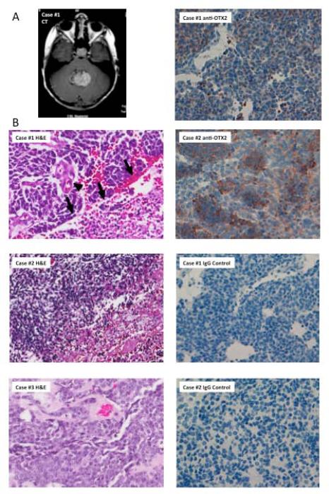
A) CT demonstrates a hyperdense 3x4 centimeter midline posterior fossa mass that appears to fill the fourth ventricle, containing punctate calcifications and cystic components. B) Haematoxylin and eosin staining of histological sections from Case #1, Case #2, and Case #3. Note that Case #1 demonstrates hyperchromatic, molded nuclei associated with vascular proliferation (arrowhead) and geographic necrosis (arrows) indicative of a severely anaplastic medulloblastoma; geographic necrosis also suggests a lack of differentiation. C) Immunohistochemical assay shows positive OTX2 expression in Case #1 (Score:40% 2+) and Case #2 (Score: 50% 2+).

