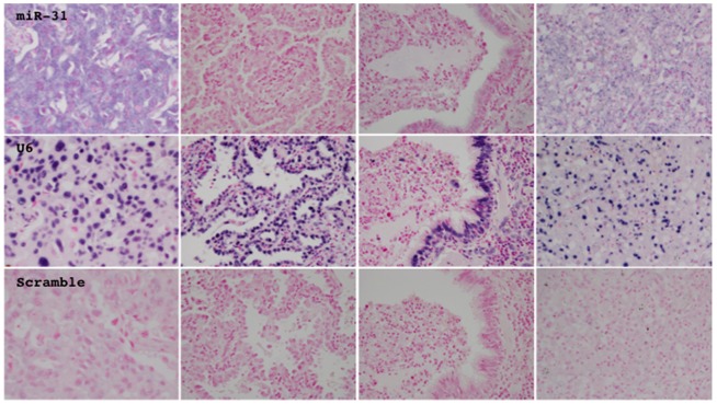
Figure 5. The expression and localization of miR-31 (top panels) and U6 snRNA (middle panels) were investigated by in situ hybridization analysis.
A probe consisting of the scrambled random sequence served as a negative control (bottom panels). Representative photographs from tumorous (left two panels) and non-tumorous tissue (right two panels), which expressed miR-31 (center two panels) or did not (lateral two panels), are presented.

