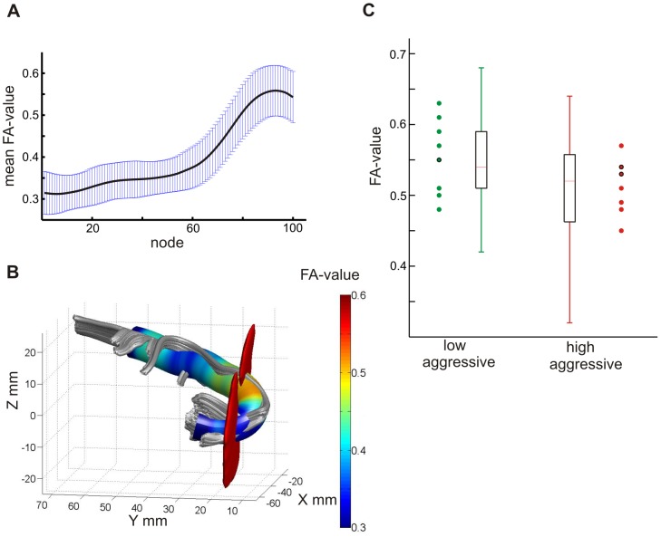
Figure 1. Descriptive results.
The distribution of mean FA-values along the left uncinate fascicle (UF) between the tract defining regions of interest (ROIs) is shown (A). Standard deviations for each of the 100 nodes are shown in blue. B shows the distribution of FA-values along the left UF for a representative subject. Tract-defining ROIs are depicted in red. C shows mean FA-values for the low and high aggressive groups as defined by the median split based on physical aggression scores for node 83 of the left uncinate fascicle. Single subject FA-values are depicted for the nine participants with the lowest and highest aggression scores. Data points encircled in black represent two cases.

