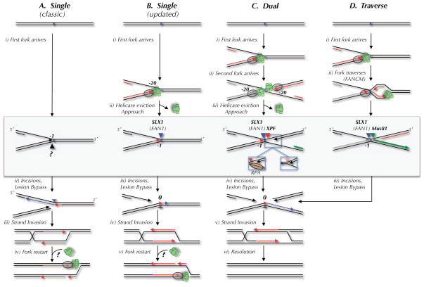
Figure 1. Possible mechanisms of replication-coupled ICL repair.
Four mechanisms of replication-dependent ICL repair are depicted. The DNA structures acted on by endonucleases in each model are highlighted by a gray box. Incisions are represented by black, blue, red, and green arrowheads. The proposed nuclease(s) that performs each incision is indicated above the arrowhead. (A) The classic ICL repair model, in which a single replication fork collides with the ICL and the leading strand template is incised (Niedernhofer et al., 2005). (B) The classic model, but taking into account the observation that leading strands initially stall 20 nucleotides from the ICL due to the MCM2-7 complex, and that incision occurs on the lagging strand template (Raschle et al., 2008). In models A and B, fork restart would require reloading of the MCM2-7 complex, for which there is no known mechanism. (C) The dual fork convergence model (Raschle et al., 2008). Left inset, 3′ incision substrate if RPA binds the lagging strand template after MCM2-7 removal. Right inset, 3′ incision substrate if parental strands re-anneal after MCM2-7 removal. (D) Traverse model (Huang et al., 2013). The only difference in the incision substrate in the dual fork and traverse models is the location of the 5′ end of the nascent strand on the right side of the ICL (green strand).

