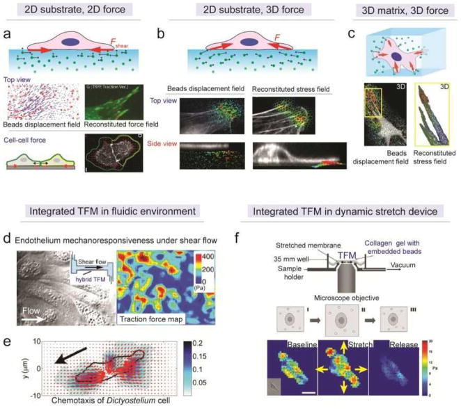
Figure 10. Microengineered traction force microscopy (TFM) in 2D and 3D contexts.
(a) (top) Schematic of 2D TFM. Although both in-plane (solid line with arrow head) and out-of-plane (dash line with arrow head) displacement occurred to each bead, only the former was recorded in conventional 2D TFM and only in-plane traction stress was reconstituted. (middle) Extracted beads displacement field and reconstituted traction force field by 2D TFM.[232] Reproduced with permission from [232]. Copyright 2008, Cell Press. (bottom) Schematic and fluorescent image showing the application of 2D TFM in characterizing cell-cell force.[228] Reproduced with permission from [228]. Copyright 2011, United States National Academy of Sciences. (b) (top) Schematic of hybrid TFM. The 3D displacement of each bead was recorded using confocal microscopy and 3D traction force on 2D substrate surface was reconstituted.[237] (middle) In-plane displacement of beads and reconstituted in-plane traction stress. (bottom) Out-of-plane displacement of beads and reconstituted out-of-plane traction stress. Reproduced with permission from [237]. Copyright 2013, United States National Academy of Sciences. (c) (top) Schematic of 3D TFM in hydrogel. (bottom) 3D displacement of beads in gel and reconstituted traction stress on 3D cell-ECM interface.[223] Reproduced with permission from [223]. Copyright 2010, Nature Publishing Group. (d) Phase contrast image (left) and traction force map (right) of a confluent endothelial monolayer under shear flow. Inset: the schematic of a circulatory fluidic channel with integrated gel substrate for performing hybrid TFM.[245] Reproduced with permission from [245]. Copyright 2012, United Stated National Academy of Sciences. (e) Cellular contractile force map of a Dictyostelium cell undergoing chemotaxis.[236] The gradient of chemokine is shown with the black arrow. Reproduced with permission from [236]. Copyright 2007, United Stated National Academy of Sciences. (f) Schematic (upper) and traction force maps (bottom) of integrated 2D TFM in a miniaturized cell-stretching device for studying the dynamics of cellular contractility during and after a transient equibiaxial stretch.[247] Reproduced with permission from [247]. Copyright 2008, Cell Press.

