
Figure 18. Integrated microenvironment for studying cancer metastasis in vitro.
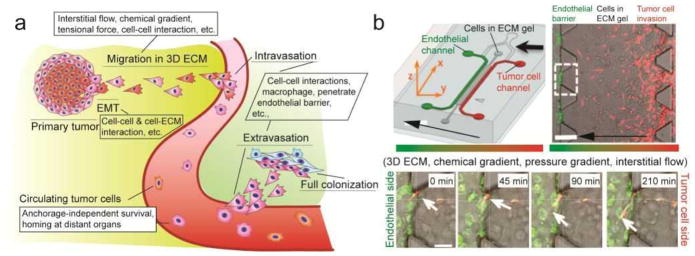
(a) Schematic of tumor cell-microenvionment interactions during the progression and metastasis of cancer. See text for detailed descriptions. (b) (upper left) Schematic of a representative gel-in-microfluidics assay for studying tumor cell intravasation. (upper right) Phase contrast image of the reconstituted tumor cell-endothelium interface. (bottom) Time-lapse confocal images showing a tumor cell invading and penetrating TNF-α stimulated endothelial monolayer.[344] Reproduced with permission from [344]. Copyright 2012, United States National Academy of Sciences.