
Figure 3. Fabricating random nanotopography.
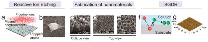
(a) Schematic of generating nanoroughness onto a substrate using reactive ion etching (RIE),[82] during which atoms were randomly and unevenly stripped off the substrate by plasma bombardment. (b) SEM image of a cancer cell on the nanorough substrate. Reproduced with permission from [82]. Copyright 2012, American Chemical Society. (c–e) SEM images of arrays of titanium dioxide (TiO2) nanotubes of different diameters, which were used to dictate differential fate of hMSCs.[83] Reproduced with permission from [83]. Copyright 2009, United States National Academy of Sciences. (f) Schematic of the spontaneous galvanic displacement reaction (SGDR), during which solute metallic ions accept electrons from substrate metallic atoms and then deposit onto the substrate as nano-scale clusters. (g) AFM image of a nanotopographical substrate created by SGDR and used for studying the response of neurons.[84] Reproduced with permission from [84]. Copyright 2010, United States National Academy of Sciences.