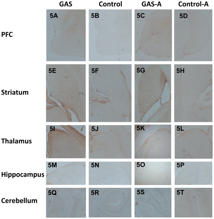
Figure 5. Immunoreactivity of IgG with the brain following GAS exposure and ampicillin treatment (low resolution).
IgG deposition in brains of GAS-exposed and control rats treated with ampicillin or water. IgG deposits in brain sections taken through the PFC (A–D), striatum (E–H), thalamus (I–L), hippocampus (M–P), and cerebellum (Q–T) of a GAS-Water (A, E, I, M, Q), Control-Water (B, F, J, N, R), GAS-ampicillin (C, G, K, O, S) and Control-ampicillin (D, H, L, P, T) rat, at a magnification ×4. Tissue sections were incubated in biotinylated-anti-rat IgG and then incubated with avidin using Vectastain ABC kit (Vector Laboratories, Burlingame, CA, USA). Anti-IgG binding to brain sections was detected using diaminobenzidine for visualization of antibody deposition. Scale bar = 200 µm.

