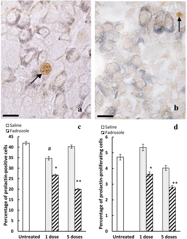
Figure 3. Effects of in vivo treatment with fadrozole on the cellular proliferation of prolactin-positive cells.
a) Double labelled immunohistochemistry for prolactin (dark blue-grey) and PCNA (brown). In untreated animals, few prolactin-positive cells are labelled for PCNA (arrow). Scale bar: 12 µm. b) After 5 doses of fadrozole it is uncommon to find cells labelled jointly for PCNA and prolactin (arrow points to PCNA- but not prolactin-positive cells). Scale bar: 12 µm. c) The percentage of prolactin-positive cells decreases significantly with respect to the untreated or control animals after 1 dose (**p<0.01 with respect to untreated animals, and p<0.05 with respect to control animals) or 5 doses (**p<0.01 with respect to untreated and control animals and p<0.05 with respect to 1 dose of the fadrozole treated animals). After 1 dose of saline the percentage decreases with respect to untreated animals (#p<0.05), but not after 5 doses. d) Fadrozole decreases the proliferation of prolactin-positive cells (*p<0.05 with respect to untreated and control animals; **p<0.01 with respect to untreated and control animals and p<0.05 with respect to 1 dose of fadrozole-treated animals).

