FIG. 5.
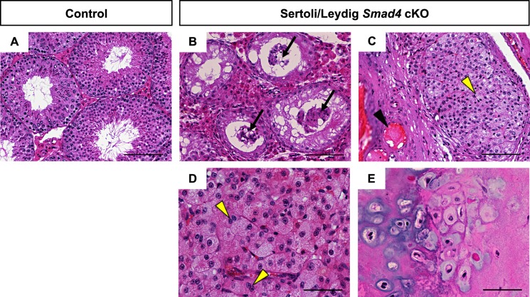
H&E stained sections from aged (56- to 62-wk old) control (A) and Sertoli/Leydig Smad4 cKO mice (B–E). Black arrows in B indicate degenerated seminiferous tubules with Sertoli cells sloughed off into the lumen. Black arrowhead in C indicates enlarged blood vessels within hemorrhagic regions. Yellow arrowheads in C and D indicate Leydig cell hyperplasia and multinucleated Leydig cells, respectively. E) Testicular teratoma containing chrondrocytes and osteoblasts was found in one of the 56-wk-old knockout testes. Bars = 100 μm (A–C) and 50 μm (D, E).

