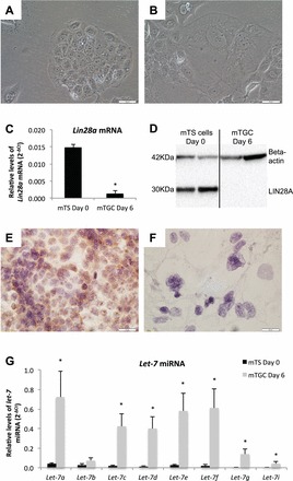
FIG. 1.

LIN28A and let-7 miRNA levels in proliferative mTS cells and differentiated mTGCs. A and B) Brightfield microscopy (20×) depicts mTS cell morphology at Day 0 under proliferative conditions, and after 6 days under differentiation conditions. C and D) LIN28A mRNA and protein levels are decreased in differentiated mTGCs compared to proliferative mTS cells. Immunohistochemistry demonstrates that LIN28A in proliferating mTS (E) is decreased when cells differentiate into mTGCs (F) after 6 days without FGF4, heparin, or conditioned medium. G) Levels of let-7 miRNA were increased in mTGCs grown under differentiating conditions for 6 days. Error bars represent SEM. Asterisk indicates P < 0.05. Bar = 0.05 mm.
