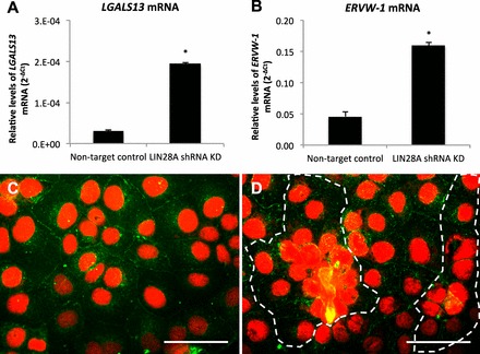
FIG. 8.

Spontaneous syncytialization in LIN28A KD in ACH3P cells. A and B) LGALS13 and ERVW-1 mRNA (qPCR) in LIN28A KD ACH-3P cells. C and D) Immunofluorescence of LIN28A KD ACH-3P cells: red, nuclei; green, plasma membrane; dashed line signifies boundary of multinucleated cell plaque. Error bars represent SEM. Asterisks indicate P < 0.01. Bar = 0.05 mm.
